HD-OPM Archive
Topics › Plaque, CAC & CT angiography

A CT angiogram shows the lumen, so it can miss early plaque inside the artery wall

Louis Nervegna · Apr 07, 2025 · ♥ 16 reactions · 25 comments · original Facebook post
Plaque, CAC & CT angiographyRisk assessment & lab tests
AI summary & key facts

Louis explains that coronary CT angiography is 'lumenography': contrast dye fills only the lumen, so early plaque growing inside the artery wall is mostly invisible until it narrows the lumen. A perfect CCTA therefore does not rule out early-stage plaque. He links a Dr Alo video on this blind spot.

I want to point something out which most people don’t seem to know.

CT-angiogram scans are examples of lumenography. These kinds of scans show the lumen of the artery. The dye is only injected into the lumen. This is the portion in contact with the circulating blood.

Plaque growth begins inside the artery wall. It only begins to impact the lumen when it gets large enough to start narrowing the area where blood can flow.

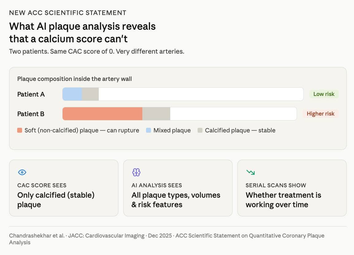
CT-angiogram scans will not show a lot of this early stage plaque inside the artery wall.
facebook.com/share/p/tK2eoLh4Be6cAo4g

A coronary CT angiogram scan with contrast (CCTA) mainly shows plaque from inside the lumen.

The lumen is the hollow space inside the artery that allows blood to flow.

A CCTA does not show all of the earlier stage plaque that is growing inside the arterial walls, unless it is extensive enough to narrow the lumen.

So you can have a perfect CCTA, but still have early stage plaque in the artery walls that hasn’t yet had an effect on the lumen.

#ctaHD

#plaque
← newer: How to follow a traditional Mediterranean diet: do's and donolder: Keto-CTA: lean keto dieters with plaque progressed; Mediterr →