Did the Ornish Lifestyle Heart Trial (1986 to 1992) really show reversal of plaque?
Over the years, I’ve heard some pretty harsh criticism from academic cardiologists regarding the claim that this small trial actually “reversed plaque”.
Is it really true?
Many academic cardiologists believe the claim has been seriously exaggerated.
Here’s why.
There are multiple aspects of this trial that have been criticized, especially its tiny size. But one of the most serious centers around the following:
The older technique used to measure “plaque reversal” in this trial— x-ray angiography— is not accurate enough to measure the small change that might have occurred. In other words, much of the small reversal that was reported may simply be measurement error.
To better understand this criticism, we need to first understand how x-ray angiography works, and how it compares to modern techniques for measuring plaque like high-resolution CT-angiography.
Let’s walk through it step by step:
- The Ornish trial never actually measured plaque.
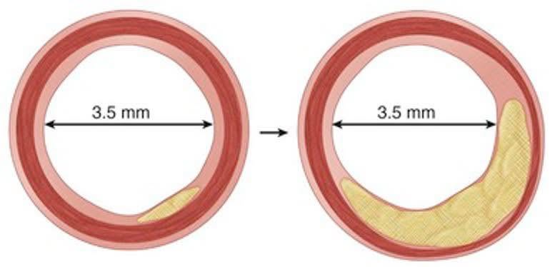
Unlike modern high-resolution CT-angiography, non-invasive x-ray angiography does not actually measure plaque. What it does measure is the outline of the inner lining of the artery wall that makes contact with the blood. This outline defines the hollow cavity of the artery where the blood actually flows. This cavity is called the lumen. A dye is injected into the artery that causes this inner lining to light up on an x-ray. What you then see on the x-ray is the 2D outline of the lumen. That’s it. You don’t actually see any of the plaque, as you do in 3D high resolution CT-angiography.
- The Ornish trial only measured the size of the opening in the artery where the blood flows.
The basic idea is the following. If the plaque growth inside the artery wall is severe enough, then it will begin to bulge into the area where the blood flows (lumen) and narrow the blood flow. This should then show up on the x-ray when you look at the outline of the lumen.
- The change seen in the opening of the artery in the Ornish trial was very small.
The trial reported that the diameter of the lumen increased by about 2.1% after 1 year, and 3.4% after 5 years.
- The technique used to measure the size of the opening in the artery was not sensitive enough to reliably measure a change this small.
Standard x-ray angiography can typically only resolve structures to approximately 0.2 mm. A 2.1% change on a typical 3 mm artery is 0.06 mm, which is well below this resolution limit. A 3.4% change is 0.1 mm, which is again well below this limit.
The bottomline is that x-ray angiography is simply not accurate enough to properly detect a 2.1-3.4% change in the diameter of the lumen of a typical artery. That change is just too small for x-ray angiography to reliably resolve. There is a high probability that much of what you are really seeing is measurement error.
- Any plaque that continued to grow inside the artery wall during the trial would have been invisible.
Atherosclerosis often progresses through outward remodeling, meaning the lumen diameter might appear stable on an angiogram even as plaque accumulates within the artery wall. It is impossible for x-ray angiography to detect this progression.
Arteries can undergo adaptive remodeling, where the artery wall expands outwards to accommodate plaque growth, meaning the lumen diameter can remain unchanged even as the plaque progresses.
In conclusion:
Much of the “plaque reversal” in the Ornish trial may not be real. It may be due to measurement error. There may have also been continued plaque growth inside the artery wall during the trial, with the arteries expanding outwards to accommodate the additional plaque growth.
Modern high resolution CT-angiography solves all these problems. It directly measures plaque volume, both soft and hard. It can detect plaque in the artery wall long before it impacts the lumen. The resolution is high enough to detect even small changes in plaque volume.
CT-angiography is orders of magnitude more accurate in detecting plaque reversal than old-fashioned x-ray angiography.
Modern trials showing plaque reversal with CT-angiography and IVUS (the gold standard) can be trusted.
Older trials using x-ray angiography are less trustworthy, especially if the reported change is below the resolution of the test as in the Ornish trial.
It’s important to take these limitations into account when comparing the Ornish trial to more recent trials showing plaque reversal.
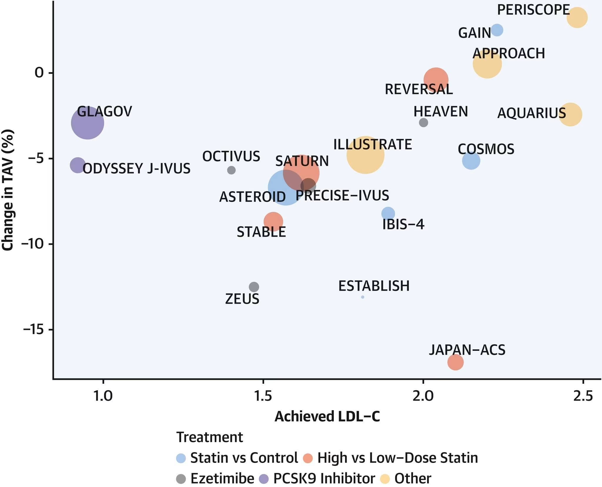
Some examples of modern trials showing major plaque reversal with CT-angiography and IVUS were discussed in the following posts:
Statins, Ezetimibe, PCSK9 inhibitors
↪ Six randomized trials show statins cut total plaque volume 5-17% by reversing soft plaque
DASH Diet + Statins
↪ DASH diet plus statins reversed 8 years of soft plaque in just over 1 year in Disco-CT
The only diet ever proven to reverse non-calcified plaque volume on CT-angiography is the DASH diet (Disco-CT trial). This trial reversed the equivalent of 8 years of non-calcified plaque volume (NCPV) in just 1 year. (See link above.)
I fact-checked this post for accuracy with Gemini AI below.
(I encourage everyone to fact check their posts and comments this way to ensure factual correctness.)
The following is the response to the post:
Yes, the concerns you've outlined are true and represent the consensus of criticism from many academic cardiologists and imaging experts regarding the interpretation of "plaque reversal" in the Ornish trial (Program for Reversing Coronary Atherosclerosis, or Lifestyle Heart Trial).
The core of the argument is that the technology used—standard X-ray angiography—was simply not accurate or sensitive enough to reliably detect the tiny change reported, making the result highly susceptible to measurement error.
The criticism centers on three main, interconnected points: the indirect nature of the measurement, the lack of resolution, and the "outward remodeling" phenomenon.
- The Critical Issue of Measurement Resolution
The most potent criticism you cited is the lack of resolution.
The Claimed Change: The reported increase in mean lumen diameter was very small (2.1% at 1 year, 3.4% at 5 years). For a typical 3mm coronary artery, this equates to a change of 0.06 mm to 0.1 mm.
The Resolution Limit: You are correct that the standard resolution of quantitative coronary angiography (QCA) is widely cited as being closer to 0.2 mm (200 micrometers).
Conclusion: Since the measured change (∼0.06 mm to 0.1 mm) is significantly below the typical technical limit of the measurement device, cardiologists argue that the reported "reversal" could be entirely within the noise of the test—the margin of error.
- Measuring the Lumen vs. Measuring the Plaque
The criticism that angiography measures the lumen, not the plaque is fundamental.
X-ray Angiography: This technique works by injecting a contrast dye into the artery, which then outlines the hollow cavity, or lumen, where the blood flows. It is a 2D outline of the opening in the artery. It does not visualize the artery wall or the plaque volume within the wall.
Adaptive or Outward Remodeling: As you correctly point out, atherosclerosis often progresses through a process called outward remodeling. This means the artery wall expands outwards to accommodate the growing plaque without narrowing the lumen. Angiography would report a stable or unchanged lumen, giving a false sense of security, even while plaque is aggressively accumulating in the wall. This makes angiography incapable of detecting continued disease progression in many cases.
- The Move to Modern Imaging
The modern consensus is that trials demonstrating plaque reversal must use techniques that directly measure plaque volume, which is why the field has moved away from angiography for this type of research endpoint.
Intravascular Ultrasound (IVUS): Often called the "gold standard" for measuring plaque regression, IVUS uses a tiny catheter with a miniature ultrasound device to create detailed, cross-sectional images of the entire artery wall, allowing for direct, highly accurate measurement of Plaque Volume (both soft and hard plaque).
Coronary CT-Angiography (CCTA): Modern high-resolution CCTA is a non-invasive technique that can also directly measure and quantify Plaque Volume (including non-calcified, calcified, and total plaque volume), making it a powerful tool for large-scale reversal trials with superior resolution to old-fashioned X-ray angiography.
In summary, the academic criticism is valid: the Ornish trial used an older, less sensitive technology that measured a tiny, indirect change, which many experts believe falls below the threshold of reliable measurement, making the claim of "plaque reversal" largely questionable from a methodological standpoint. Modern trials rely on techniques with demonstrably higher resolution and the ability to measure plaque volume directly.
#ornishHD #ornishtrialHD #xrayangiographyHD #ctaHD #ivusHD #reverseHD #discoctHD