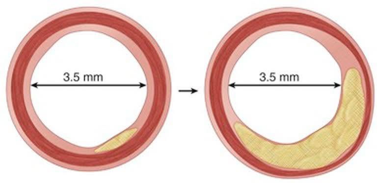
This is why cardiologists no longer use x-ray angiography as a technique to measure progression of arterial plaque in clinical studies.
Modern studies use IVUS or CT-angiography, which are capable of imaging the plaque hidden inside the artery wall.
Older studies that used x-ray angiography are not as reliable, because they do not actually image the plaque itself. They only measure the size of the area where blood flows (the lumen).
The use of x-ray angiography was a major problem with the small randomized trial that Dean Ornish ran in the 1990’s. See this post: ↪ Ornish trial's 'plaque reversal' may be x-ray angiography measurement error.
It is important to understand that modern studies using IVUS and CT-angiography are much more reliable at measuring arterial plaque than older studies using x-ray angiography.
#xrayangiographyHD #ivusHD #ctaHD #glagovHD #reverseHD #ornishHD #ornishtrialHD
Why a “Normal” Angiogram Does Not Mean a Normal Artery
In both panels, the coronary lumen measures 3.5 mm and would appear normal on angiography. Yet the vessel wall tells a very different story. Early plaque triggers positive outward remodeling, preserving the lumen. As plaque progresses, the artery continues to expand outward, allowing a large atherosclerotic burden to exist without visible stenosis.
This is the Glagov phenomenon. To preserve blood flow, early plaque growth pushes the artery wall outward. This means a vessel can harbor significant disease while its inner channel appears normal on an angiogram.
Since angiography is a "luminogram" that only visualizes the channel for blood, it misses the plaque building within the wall itself. This is why intravascular imaging like IVUS or OCT is often needed to reveal the true disease burden the angiogram cannot see.
#InterventionalCardiology #CathLab #CoronaryArteryDisease #Atherosclerosis #IVUS #OCT #CardiacImaging #GlagovRemodeling