Results of the YELLOW-III Trial
In just 6 months time, patients taking a combination of high dose statins + pcsk9 inhibitors saw a major strengthening of the fibrous cap that protects existing plaque, and a major reduction in total plaque volume.
The LDL of participants in this trial was in the 30-40 mg/dL range.
The trial has been described as “melting away plaque like butter”, because of the dramatic reduction seen in the soft lipid core (the “butter”) inside the plaques. See this previous post:
facebook.com/share/v/1CHZCYr1jK
Fibrous cap thickness (FCT) increased by over 30%. This was measured using invasive optical coherence tomography (OCT), which is the gold standard for measuring FCT.
Total plaque volume (PAV) decreased by an absolute reduction of 1.38%, as measured by IVUS. This was driven primarily by reduction of the soft lipid core inside the plaque. IVUS is the gold standard for imaging plaque. An ultrasound camera is inserted invasively into the arteries during a coronary angiogram. An absolute PAV reduction this large represents a reversal of multiple years of progression, in just months time.
PAV is a measure of total plaque volume. It measures the sum of all plaque, both soft and hard. The units of PAV are in percent. PAV measures the percentage of the total scanned vessel volume that is occupied by plaque. For example, a PAV of 15% would mean that 15% of the volume of scanned arteries would be composed of plaque, while the remaining 85% would be normal tissue.
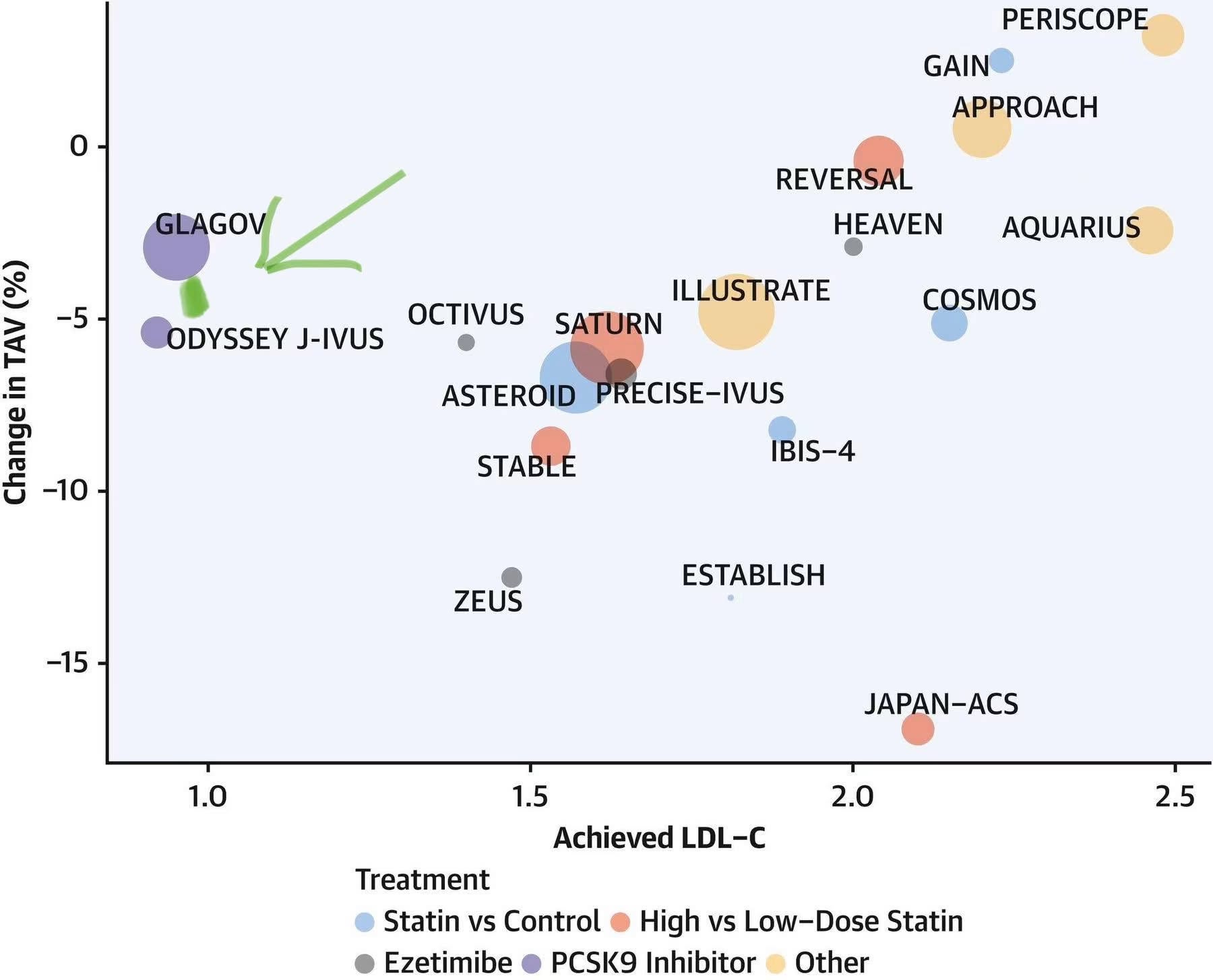
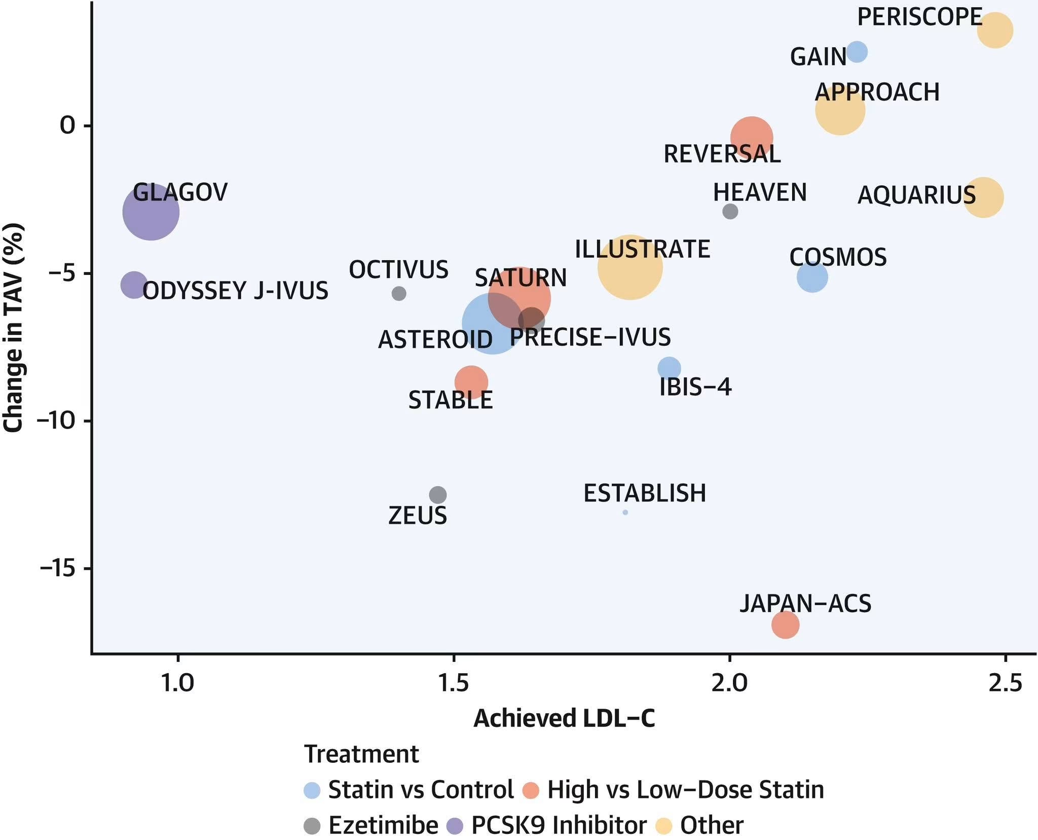
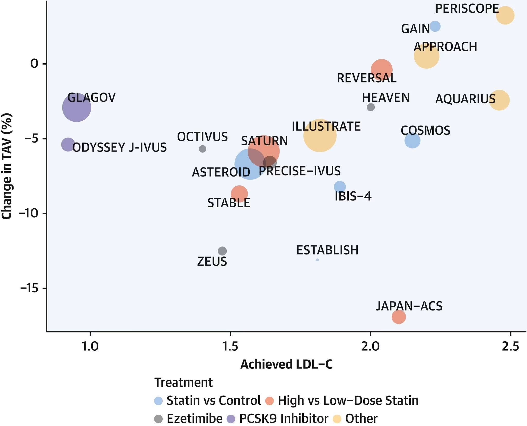
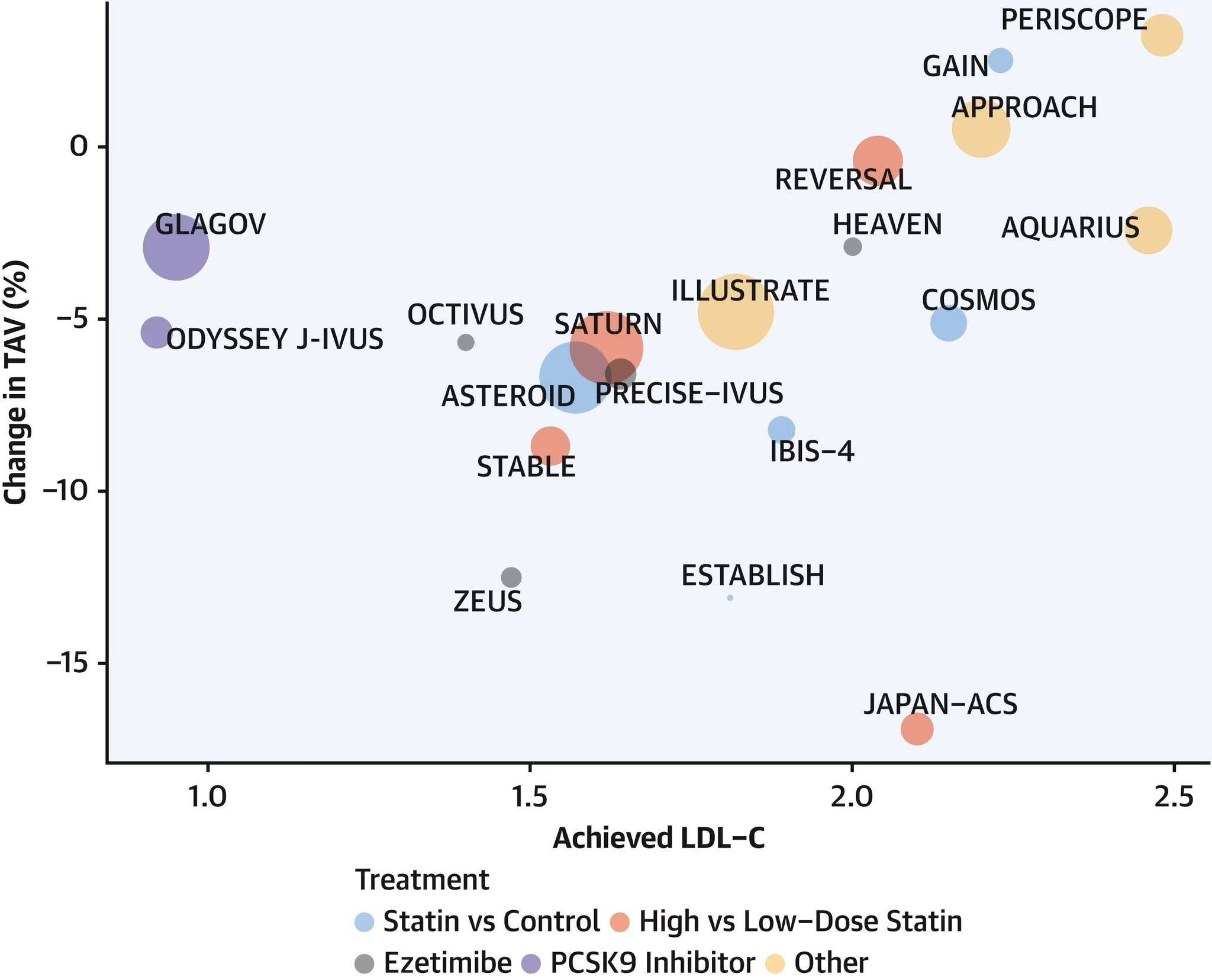
PAV above 15% is often considered Stage 3 (Severe) plaque burden. While a universal single-name scale does not exist, this classification is part of coronary CTA plaque volume severity stages (or AI-QCTCAD staging), often categorizing >15% PAV as Stage 3 (Severe). So a 1.38% absolute reduction from, say, 15% down to 13.62% in just 6 months is a very big deal. We know from previous trials like GLAGOV and NISSEN that every 1% reversal of PAV is associated with a roughly 20% reduction in major adverse cardiac events (MACE).
It is the very short time frame that is most remarkable about this study!
See this post below for a comparison of the total plaque reduction in YELLOW-III to previous trials:
↪ YELLOW-III reversed total atheroma volume 4.4% in 6 months at LDL of about 39 mg/dL
The researchers also demonstrated that the reversal and stabilization of plaque on imaging was accompanied by improvements in the underlying pathology, including restored mitochondrial function, suppressed inflammation, and alleviated oxidative stress.
Link to press release from Mount Sinai Hospital:
reports.mountsinai.orgCardio2024 yellow trialreports.mountsinai.orgLink to full study:
Paper · JACC Cardiovasc Imaging · 2026Intracoronary Imaging and Transcriptomic Characteristics Before and After Maximum Lipid-Lowering Therapy: YELLOW-III Study of Evolocumab and Maximum Statin Therapy in Chronic Coronary Disease.Conclusion: In stable coronary artery disease, intravascular imaging-verified beneficial changes in plaque morphology after evolocumab treatment were associated with restored mitochondrial function, suppressed inflammation, and alleviated oxidative stress. Similar studies of plaque morphology with noninvasive modalities would allow the confirmation of novel pathways of plaque stabilization in response to maximized…pubmed.ncbi.nlm.nih.govQuoted from the press release:
The study, known as Yellow III, used advanced multimodality imaging to show favorable plaque characteristics after a 26-week regimen of evolocumab, including substantial reductions in total cholesterol, LDL cholesterol, and total/HDL cholesterol ratios. More specifically, the investigation showed a significant increase in the minimum fibrous cap thickness (FCT) through optical coherence tomography (OCT), reduction in lipid core burden index at the maximal 4-mm segment (maxLCBI4mm) through near-infrared spectroscopy [NIRS], and reduction in atheroma volume through intravascular ultrasound in angiographically nonobstructive lesions.
In the Yellow III trial, 137 patients scheduled for elective coronary angiography were prescribed maximum-dosage statin therapy for at least four weeks before undergoing multimodality intracoronary imaging. They were then given evolocumab (140 mg) every two weeks for 26 weeks and reimaged to assess changes in plaque morphology and composition.
By using all three modalities [IVUS, OCT, NIRS] for the first time in a study of this type we were able to demonstrate a measurable improvement in fibrous cap thickness, as well as in plaque volume,” says Dr. Kini, Zena and Michael A. Wiener Professor of Medicine (Cardiology) at the Icahn School of Medicine at Mount Sinai. “In addition, blood samples were drawn to enable us to conduct a gene expression analysis of peripheral blood mononuclear cells. This will help us uncover through ongoing research the molecular mechanisms responsible for beneficial changes in atherosclerotic lesions of patients treated with evolocumab.”
Quoted from the full study:
A total of 110 stable coronary artery disease patients receiving maximally tolerated statin therapy were treated with evolocumab 140 mg every 2 weeks for 26 weeks. Serial optical coherence tomography, intravascular ultrasound, and near-infrared spectroscopy of nonobstructive lesions were performed in all patients at baseline and 26-week follow-up.
After evolocumab treatment, optical coherence tomography-verified fibrous cap thickness (FCT) increased from 70.9 ± 21.7 to 97.7 ± 31.1 μm (mean 26.8 ± 22.3 μm; P < 0.001), and maximal lipid core burden index within 4 mm (LCBI) decreased from 306.8 ± 177.6 to 213.1 ± 168.0 (mean -93.7 ± 140.5; P < 0.001). In addition, there was a significant reduction in the percentage atheroma volume (-1.38% ± 1.48%; P < 0.001) defined by intravascular ultrasound.
Among 110 patients, 88 (80%) demonstrated FCT increase >0 (FCT responders), and 86 (78%) demonstrated LCBI reduction >0 (LCBI responders). Whereas multiple canonical pathways modulating immune cell adhesion, recruitment and communication were down-regulated, those associated with mitochondrial function, cell survival, and protein synthesis were significantly up-regulated in response to evolocumab.
In stable coronary artery disease, intravascular imaging-verified beneficial changes in plaque morphology after evolocumab treatment were associated with restored mitochondrial function, suppressed inflammation, and alleviated oxidative stress.
#reverseHD #statinreverseHD #pcsk9reverseHD #pavHD #plaqueHD #ivusHD #lipidcoreHD #fibrouscapHD #statinsHD #pcsk9HD #yellowiiiHD #meltingbuttersignHD #fctHD #octHD